Real-time imaging is very much essential to spot abnormalities and evaluates tissues and organ health during check-ups. Hence, a precise and reliable ultrasound is necessary for patients who seek medical advice and diagnosis.
Vinita Hospital utilizes the most adaptable ultrasound scan in Chennai that are easily accessible and can be technically used for imaging each and every part of our body. The ultrasound preliminary reports allow the clinical staff to detect the critical findings and a final report is prepared by the radiologist thereafter. The patients can get very high-resolution images due to the advanced ultrasound machine.
Moreover, ultrasound services are provided for pelvic, obstetrics, prostate, regional, abdominal, 3D/4D obstetrics, cranial, neck, scrotal, thyroid, and musculoskeletal with an affordable ultrasound scan price in Chennai.
What is an Ultrasound? (Ultrasound Scan)
An ultrasound is an imaging test that makes use of sound waves to create a (sonogram) picture of organs, tissues, and other structures inside the body. Ultrasounds do not use any radiation, unlike X-rays. An ultrasound can also show the body parts in motion, such as a heartbeat or blood flow through blood vessels.
It is generally non-invasive. Common reasons for an ultrasound scan in Chennai include investigations of an individual’s abdominal and pelvic organs, vascular and musculoskeletal systems, and to check fetal development during pregnancy.
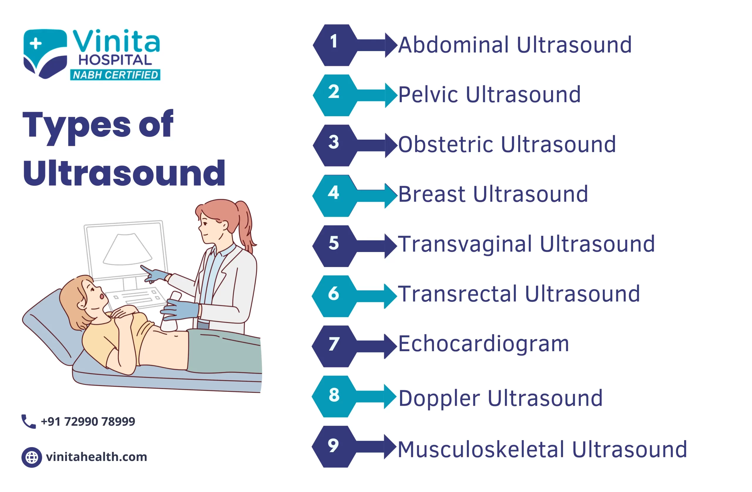
There are two main categories of ultrasounds: diagnostic ultrasound and pregnancy ultrasound.
Diagnostic Ultrasound is used to view and give information about other internal parts of the body. These include the blood vessels, heart, liver, kidneys, bladder, and female reproductive organs.
Pregnancy Ultrasound is used to know the details of an unborn baby. This ultrasound scan in Chennai provides information about a baby’s development, growth, and overall health.
An ultrasound can be used in several ways, depending on the type of ultrasound besides which part of the body is being checked.
Diagnostic ultrasound may be used to:
Find out if the blood flows at a normal rate and level.
Check if there is an issue with the heart structure.
Check for blockages in the gallbladder.
Check the thyroid gland for non-cancerous or cancer growth.
Check for abnormalities in the pelvis, abdomen, and kidneys.
Help guide a biopsy procedure.
In women, diagnostic ultrasound scanning may be used to:
Check for breast lumps to see if they might be cancer. (This ultrasound scan in Chennai is also used to check for breast cancer in men, but compared to men, women are more prone to breast cancer).
Helps in finding the exact cause of pelvic pain, and abnormal menstrual bleeding.
Help diagnose or monitor infertility treatments.
In men, diagnostic ultrasound scanning may be used to diagnose disorders of the prostate gland.
A pregnancy ultrasound may be used to:
Confirm pregnancy.
Check the position as well as the size of the unborn baby.
Check to see if you are pregnant with more than one baby.
Estimate the duration of how long you have been pregnant. This is termed gestational age.
Check for signs of Down syndrome, which might include thickening in the back of the baby’s neck.
Check for congenital birth defects in the spinal cord, brain, heart, or other parts of the body.
Check the amount of amniotic fluid (a clear liquid that surrounds an unborn baby during pregnancy to protect the baby from outside injury and cold. It also aids in promoting bone growth and lung development).
The comprehensive facilities provided at Vinita Hospital are:
The latest technology ultrasound scanning is provided with reasonable ultrasound scan price in Chennai.
3D/4D Ultrasound of abdomen, obstetrics, and gynaecological cases.
Musculoskeletal ultrasound for elbow, wrist, shoulder, knee, ankle, hip, etc.
Paediatric ultrasound which includes the abdomen, pelvis, hip, neuro sonograms etc.
Vascular ultrasound that includes Colour Doppler of the upper/lower extremities, abdomen scrotal, carotids, penile, gynaecology/ obstetrics etc.
Transrectal ultrasound scan in Chennai for prostate, rectum etc.
Technical expertise is patient-centric to provide an accurate diagnosis.
Spontaneous access to treatment by health care providers with quick reports.
Patients experience minimal discomfort throughout the Procedure.
A multi-disciplinary team is available for opinion and management.
Following the ultrasound scan in Chennai, your doctor at Vinita Hospital will see the images and check for any abnormalities. You may also need to undergo other diagnostic techniques, such as an MRI, CT scan, or a biopsy depending on the area examined.
If your healthcare provider is able to make a diagnosis depending on your condition based on the reports of ultrasound, they will begin your treatment immediately.
Overview
Types
Uses
Facilities
Our Doctors & Specialties
Frequently Asked Questions
Can we eat before ultrasound?
It depends on the type of ultrasound being performed. For some types of ultrasound, such as abdominal or pelvic ultrasound, you may be asked to fast for a certain period of time before the scan. This is because the presence of food or drink in the digestive system can interfere with the quality of the images.
How long do ultrasound results take?
The time it takes to receive ultrasound results can vary depending on a few factors, such as the complexity of the scan and the healthcare provider’s workload. In most cases, the radiologist or physician who interprets the ultrasound will send a report to the healthcare provider who ordered the test within 24 to 48 hours.
What is the most common ultrasound scan?
The most common ultrasound scan is the obstetric ultrasound, which is used to evaluate the health and development of a fetus during pregnancy. Obstetric ultrasounds can be performed at various stages of pregnancy, and they are used to confirm pregnancy, estimate the due date, evaluate the fetus’s growth and position, check for any abnormalities or birth defects, and monitor the health of the mother’s reproductive organs.
Why would I need an ultrasound scan?
You may need an ultrasound scan for several reasons. Here are some common indications for an ultrasound scan:
Diagnostic purposes: Ultrasound scans are frequently used to diagnose various medical conditions. They can help evaluate the health and function of organs such as the liver, gallbladder, kidneys, spleen, thyroid, and reproductive organs. Ultrasound scans can detect abnormalities, such as tumors, cysts, stones, or inflammation, providing valuable diagnostic information.
Pregnancy monitoring: Ultrasound scans are an essential tool for monitoring fetal development during pregnancy. They can determine the age of the fetus, check for multiple pregnancies, assess the growth and position of the fetus, and detect any potential abnormalities.
Abdominal pain or discomfort: If you experience abdominal pain or discomfort, an ultrasound scan can help identify the underlying cause. It can detect conditions such as gallstones, kidney stones, appendicitis, abdominal hernias, or liver diseases.
Pelvic pain or menstrual irregularities: Ultrasound scans can be used to examine the pelvic region in both males and females. They can help diagnose conditions such as ovarian cysts, uterine fibroids, pelvic inflammatory disease, or prostate enlargement.
Evaluation of blood vessels: Ultrasound scans can assess blood flow and detect abnormalities in the blood vessels. They are commonly used to evaluate conditions such as deep vein thrombosis (DVT), carotid artery disease, peripheral artery disease (PAD), or varicose veins.
How is an ultrasound scan performed?
During an ultrasound scan, the following steps are typically involved:
Preparation: Depending on the area of the body being examined, you may be asked to change into a hospital gown or remove certain clothing and jewelry that may interfere with the scan. In some cases, you may be required to follow specific instructions, such as fasting or drinking water to fill your bladder, if applicable to the area being scanned.
Positioning: You will be positioned on an examination table, either lying down or sitting, based on the part of the body being scanned. The sonographer (ultrasound technician) will ensure your comfort and proper positioning to obtain the best possible images.
Gel application: A clear, water-based gel will be applied to your skin over the area being examined. This gel helps transmit the sound waves and allows the transducer to glide smoothly over your skin.
Transducer placement: The sonographer will hold a handheld device called a transducer. The transducer emits high-frequency sound waves into your body and also receives the echoes of those sound waves bouncing back from your internal organs.
Image capture: The sonographer will move the transducer gently over the area of interest, applying slight pressure as needed to obtain clear images. They may ask you to hold your breath briefly or change positions to enhance visualization of specific structures.